Monday, January 7, 2019

Algae [Plant kingdom]

ALGAE

  • M.O.P. Iyengar[1886-1963] was born in Madras.
  • He started his Research in phycology especially about volvocales.
  • The great algologist Fritsch was the guide of M.O.P. lyengar for algal studies at London.
  • Term "Algae" was given by Linnaeus.
  • Phycology - Study of algae.
  • Father of Phycologoy - Fritsch  Book  "Structure & Reproduction of algae"
  • Algae are found in both fresh and marine water.
  • Algae are found in many forms like filamentous, colonial.
  • Algae are surrounded by mucilaginous sheath and below the sheath cell wall is present which is made up of cellulose and pectin but mainly made up of cellulose, galactans, mannans and minerals like calcium carbonate.
  • On the basis of structure, algae are thalloid i.e. plant body is not differentiated into root, stem and leaves. vascular and mechanical tissue system is also in algae.
  • On the basis of nutrition,algae are photo-autotrophic. They have chloroplast in which photosynthetic pigments are present. classification of algae is mainly based on pigments. Chl-a, carotene are universal pigments of algae.
  • Life cycle is mainly haplontic due to zygotic meiosis.

Reproduction

1. Vegetative  reproduction  

  • Binary fission - Cell is divided into two parts and nucleus is also divided into two parts by mitosis. eg . Found only in unicellular algae 
  • Fragmentation - Filaments break down into small pieces & form new filaments. eg. All filamentous algae.

2. Asexual reproduction  :

It is a common method of  reproduction. 

3. Sexual reproduction :

  • Male sex organ is called antheridium and female is called oogonium. The sex organs of algae are unicellular  & jacketless. But exceptionally sex organs of  green algae Chara (Chara - green algae - known as stone wort ) are muticellular and jacketed.
  • The male sex organ of chara is known as  globule  and female is known as  nucule.
  • plant body of algae is haploid so sexual reproduction take place through zygotic meiosis. So their life cycle is  haplontic. But exceptionally brown algae are diploid so that sexual reproduction take place through gametic meiosis in them. so their life cycle is diplohaplontic
  • Algae reproduce by zygotic meiosis i.e. first division in zygote is meiosis so embryo is not formed.

Sexual Reproduction is of three Types

  • (a)    Isogamous          -    Chlamydomonas debaryabnum, Ulothrix, Ectocarpus 
  • (b)    Anisogamous     -     Chlamydomonas braunii 
  • (c)    Oogamous         -     Chlamydomonas  coccifera, Sargassum, Volvox.

Saturday, June 30, 2018

Kingdom Fungi


  • Father of mycology- Pier' Antonio Michelli.
  • Father of Systematic mycology - E. M. Fries .
  • Father of modern mycology and plant pathology- H. A. de Bary.
  • Father of indian mycology and plant pathology - E. J. Butler.
  • K. R. Kirtiker was first indian scientist who collected and identified Fungi.

Distribution

 There are more than 100,000 species of fungi, which are cosmopolitan in distribution.


Habitat:

 Fungi are ubiquitous i.e. found almost everywhere. They flourish well in moist, dark and warm habitat. The most usual habitat of Fungi is wet soil rich in humus. A few forms are aquatic (e.g., Saprolegonia, Allomyces, Achyla ).
Nutrition

  • Fungi lack chlorophyll and are unable to synthesize their own food by photosynthesis. Therefore, they obtain their nutrition from the external source by absorption and by the process of extra cellular digestion. Such mode of nutrition is called Saprophytic mode of nutrition.
  • Some fungi are symbiotically associated with algae to form Lichens. A few forms symbiotic association with the roots of higher plants (e.g., Pinus) known as mycorrhiza.

Thallus Organization

  • The plant body of fungi is thallus which may be acellular or multicellular. Acellular thallus may be motile (e.g., Synchytrium ) or non-motile (e.g., Saccharomyces).  Multicellular thallus is tubular, filamentous, branched and is called mycelium. The unit is of mycelium is hyphae.
  • The mycelium may be aseptate and septate. Aseptate mycelium lacks septa. It is multinucleate and is called Coenocytic, e.g., Phycomycetes. In aseptate mycelium, septa formation occurs at the time of injury and during separation of reproductive structures. Septate mycelium is partitioned into separate compartments by means of cross walls(septa). Individual cells may be uninucleate, Binucleate and multinucleate.
  • Each septum is perforated by a central pore. The pore may be simple or dolipore[barrel-shaped swelling around their central pore].
  • Mycelium may be eucarpic[only a part forms reproductive body] or holocarpic[the whole mycelium is transformed into reproductive body].

Cell Structure

  • Fungi are eukaryotic and posses true nuclei. Fungal cells are bounded by definite cell wall. The cell wall is made up of Chitin/ Fungal cellulose[nitrogen containing polysaccharide or heteropolymer of acetyl glucosamine which is also found in insects]. True cellulose is found in cell wall of some Phycomycetes (e.g., Phytophthora).
  • The cell wall encloses protoplast, which is differentiated into plasma membrane, cytoplasm, nucleus and vacuoles. Cytoplasm has all the eukaryotic cell organelles, except plastids. The dictyosomes are also not typical. Lomasome is a convoluted complex of membranous outgrowth of plasmalemma.
  • Vacuoles are many but small.
  • Nuclei are smaller as compared to those of higher plants. Nuclei undergo intranuclear spindle formation[karyokinesis, Moore, 1961].
  • Food reserve is glycogen[animal starch] and oils.

Reproduction

  • Reproduction can be vegetative, asexual and sexual.

Vegetative Reproduction

Vegetative reproduction occurs by the following methods:-
  1. Fragmentation :- Mycelium may break accidentally or due to decay into two or more parts. each segment develops into complete mycelium.
  2. Fission :- It occurs into unicellular forms where cell division produces two daughter cells. e.g., Yeasts. 
  3. Budding :- The protrusion grows out into a bud. The bud constricts at the base and separates into new individuals e.g., yeast. Sometime a number of buds may be seed attached to the parent cell[Torula stage].
  4. Chlamydospores :-  They are thick walled, black, multinucleate resting spores formed by the collection of protoplasm at one or many places. They are mostly intercalary and after the separation, germinate to produce new mycelia.e.g., Rhizopus, Mucor, Ustilago etc.
  5. Oidia/Arthrospores:- Oidia are thin walled structures produced in chain. They are formed by the segmentation of hypha in presence of excess water, sugar and salts. They germinate immediately after liberation and produce new mycelia, e.g., Mucor. The Oidia formation in Mucor represents Torula stage.

Asexual reproduction

  • Asexual reproduction is accomplished by means of asexual spores formed by mitosis, so also called mitospores. Asexual spores are of two types, motile[zoospores] and non-motile[aplanospores].

Zoospores

  • The zoospores are thin walled flagellate spores produced inside a sac like structure known as zoosporangium. Zoospores may be uniflagellate or biflagellate. The flagella occur anteriorly, laterally or posteriorly, e.g., aquatic fungi.

Aplanospores

  • Aplanospores are of two types, sporangiospores and conidia.
  • Sporangiospores are thin walled, non-motile, uni or multinucleate spores produced endogenously in sporangium. After dispersal and germination they produce new mycelia, e.g., Rhizopus, Mucor etc.
  • Conidia(Gr. konis- dust, idion - diminutive) are non-motile, thick walled asexual spores produced exogenously at the tip of special hyphae, called conidiophores in a basipetal succession. Conidia are dispersed by wind. On falling on suitable substratum, each conidium produces a new mycelium e.g., Penicillium, Aspergillus etc.

Sexual Reproduction

  • Most true fungi reproduce by sexual method, but in the members of the fungi imperfecti (Deuteromycetes), the sexual stage is either absent or unknown. The sexual reproduction in true fungi is affected by the fusion of two nuclei of different parentage, which may be carried in motile or non-motile gametes, in gametangia, or insomatic cells of the thallus.

Types of sexual reproduction

  • There are different types of sexual reproduction in different groups of fungi.

Sex organs 

  • There are two types of organs(gametangia) male and female. The male sex organ is called anthredium while the female sex organ is called oogonium(in the members of Oomycetes) or ascogonium( in the members of Ascomycetes).There is no development of sex organs in the members of Basidiomycetes, though they reproduce sexually  by somatogamy. There is no sexual reproduction at all in the members of Deuteromycetes, so they are called fungi imperfecti.

Classification of Fungi

  • A number of criteria are used for classification of fungi. The important ones are morphology of vegetative structures, morphology of reproductive structures, types of meiospores and mitospores, life cycle, physiology, biochemistry etc.

Wednesday, June 6, 2018

Photosynthesis in Higher Plants

Photosynthesis in Higher plants pdf notes Click on the link mentioned below to read now
Photosynthesis in Higher Plants

Follow out blog so that you'll notified each and every time whenever we post something new.
Thanks.. Please do share with your friends also.

Saturday, March 17, 2018

Phylum Platyhelmithes - The Flat worms

Gegenbeur (1859) placed flat worms in a separate group and gave the term Platyhelmithes. The Phylum Platyhelminthes includes about 13,000 species.

General Characterstics

  1. Habitat: The flatworms are mostly parasites but some flatworms are free-living.
  2. Body Form: The body is dorso-ventrally flattened and is without true segments.
  3. Symmetry: bilaterally symmetrical.
  4. Germ layers: From the evolution point of view, they are first triploblastic animals, e.g., consisting of three germ layers: ectoderm, mesoderm and endoderm.
  5. Level of Organisation: The flatworms have organ level of organisation.
  6. Body wall: The body covering is soft and may or may not be ciliated. Rod shaped bodies, the rhabdites are present in epidermal cells of the living platyhelminthes. They are protective in nature and used for capturing food.
  7. Body cavity: The space between the body wall and the organs are filled with a peculiar connective tissue, called the parenchyma. The parenchyma helps in transporting the food materials. Flat worms are acoelomates.
  8. Digestive Tract: If present, is incomplete ( without anus ) . Digestive tract is absent in Tapeworms.
  9. Skeletal, Respiratory and Circulatory system are absent. The fluid in the parenchymal network maintains the body shape. It is called hydoskeleton.
  10. Excretory system : It consists of special flame cells ( Solenocytes / Protonephridia) which are meant for excretion and osmoregulation.
  11. Nervous System : The nervous system is ladder like. It consists of the brain and two main longitudinal nerve cords connected at intervals by transverse commissures.
  12. Reproductive System : These animals are generally hermaphrodite[i.e., male and female organs in single individual] , and the reproductive organs are well developed. The fertilization is cross and internal. In tapeworms self fertilization is found. 
  13. Development : Life cycle is complicated in most flatworms with one or more larval stages. In liver fluke miracidium, sporocyst, radia, cercaria and metacercaria larvae are present.
  14. Regeneration : It is well marked in some flatworms like Planaria. 

Wednesday, November 1, 2017

FEMALE REPRODUCTIVE SYSTEM

  • Consists of pair of ovaries, a pair of oviducts(Fallopian tubes), a sac-like uterus and the external genitalia like vulva and genital glands.

Ovaries

  • Almond shaped structures in the upper part of the pelvis. It lies in the lower part of abdomen attached to dorsal wall by mesovarium near kidney. Attached to uterus by ovarian ligament.
  • Each ovary measures about 3cm*2cm*1cm surrounded by the fold of peritonium.
  • Blood vessels and nerves enters through a narrow conducting part, hilus having continuity with mesovarium. 
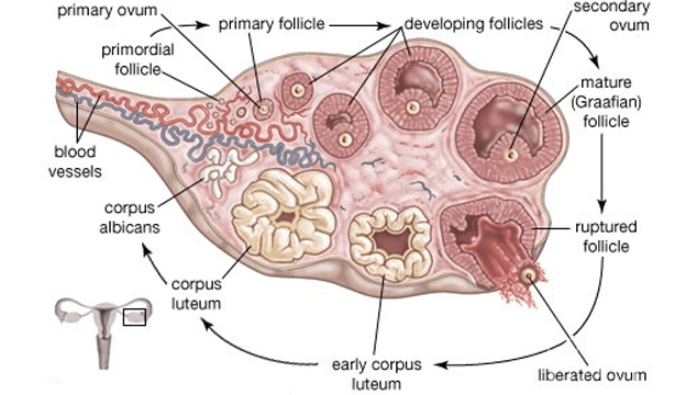
  • In wall of ovary visceral peritonium, germinal epithelium and tunica albuginea are present. Germinal epithelium is made up of Primary Germ Cells(PGC) .
  • Ground part of ovary is stroma differentiated into outer cortex and inner medulla , mesodermal in origin.
  • P.G.C. of germinal epithelium are endodermal in origin.
  • First of all from P.G.C., follicle cells are formed which fuse to form egg nest in which one is developing ovum, rest of the cells are destroyed to give nourishment to developing ovum.
  • Destroyed cells atractic cells. The phenomenon is called atrasia.
  • First it is single layered termed as primary follicle. Soon it becomes double layered i.e., secondary follicle after that it is converted into graffian follicle or mature follicle.
  • Its formation is folliculogenesis under control of FSH and LH.

Accessory Organs

  • The oviduct, uterus, vagina , clitoris, the accessory glands and mammary glands constitutes the secondary sexual organs of human female.

Fallopian Tubes/Oviducts

Fallopian tubes are a pair of muscular tubes(10cm) that extend laterally from the uterus on either side of the wall of pelvis. Fertilization occurs into it.
It is supported by double fold of peritonium.
It shows 4 regions- 
   (i) Infundibulum-
  • Infundibulum has broad, funnel shaped proximal part.
  • Its margin bears fimbriae which has finger like projections.
  • In funnel ostium aperture present, from it eggs enter into duct.
  (ii) Ampulla-
  • Ampulla is a thin walled, longest part lined with ciliated epithelium and is the site for fertilization.
  (iii) Isthmus-
  • Very short, narrow thick walled straight part.
 (iv) Uterine part-
  • It is a narrow part that communicates with uterine cavity.
  • Tubectomy (or Tuballigation , the cutting or ligation of fallopian tube prevents the entry of ovum into it.

Uterus(Womb)

  • For viviparous development in mammal it makes the house of embryo.
  • In woman it is sac-like, thick walled, pear shaped, muscular and glandular part of 8*5*2 cm in size.
  • It is large, pyriform, highly elastic and development of embryo takes place into it.
  • It is located above and behind the urinary bladder and is attached to body wall by mesovarium ligament.
  • It shows four regions-
  1. Upper wide, dome shaped fundus that receives fallopian tubes.
  2. Cornuae the upper corners where the oviducts enter into the uterus.
  3. Middle large body or corpus which is the main part.
  4. Lower narrow cervix that projects into the vagina.
  • In the wall of uterus outer layer is perimetrium, middle layer is myometrium and inner is endometrium.
  • Myometrium consists of inner and outer of longitudinal smooth muscles and middle circular muscles. Longest involuntary muscles present here.
  • Endometrium consists of inner epithelial layer and underlying connective tissue layer(lamina properia) with many coiled tubular glands and screw-like blood vessels. This undergoes cyclic changes during menstruation.
  • Cervix has two openings, os internalis towards corpus and os externalis towards vagina.
  • Cervix communicates with uterus by internal os. and with vagina by external os.
  • Cavity of cervix between external and internal os. is cervical canal.
  • Cervix is composed of  the biggest and most powerful sphincter muscles in the body It is strong enough to hold about 7 kg of tissue fluid.
  • After implantation and pregnancy placenta is formed by the contribution of uterine wall.
  • Hysterectomy is the surgical removal of uterus due to defect.

Vagina

  • A canal below uterus opens to outside between urethral pore and anus or in the urino-genital sinus(rat).
  • Median, elastic, muscular tube 7.5 cm long. Opens into vestibule by vaginal orifice.
  • It receives penis during copulation and also serves as birth canal.
  • Space between vaginal wall and cervix is fornix.
  • Initially its opening remains covered with a membranous hymen which ruptures either during sexual intercourse or due to any other vigorous physical exercise or work.
  • A slit in hymen allows menstrual flow to pass out.

External Genitalia

  • The external part around the vaginal pore is vulva with upper pad-like part, mons pubis with hairs.
  • The middle cut the,vestibule, is laterally bound to with two pairs of lips(skin folds), outer labia majora and inner labia minora.
  • At the anterior junction of labia minora a small erectile clitoris present, homologous to penis.
  • In the vestibule the urethral opening is present on the upper side and vaginal orifice at the lower side.
  • Area between fourchette and anus is perineum.

Glands

Vestibular glands are of two types:

  1. Lesser Vestibular glands/Paraurethral glands/Glands of skene:  Numerous minute glands that are present around the urethral orifice. These glands are homologous to the male prostrate and secrete mucous.
  2. Greater Vestibular glands/Bartholin's glands: Paired glands situated one on each side of the vaginal orifice. These glands are homologous to bulbourethral glands/ cowper's glands of male and secrete viscous fluid that supplements lubrication during sexual intercourse. 

Breasts

  • One pair, their development is under control of pituitary.
  • Nipples present (absent in prototheria). Around nipples area mammary glands present, in it melanin is maximum.
  • Milk is synthesized by lactogenesis under control of prolactin. Milk is secreted under control of oxytocin.
  • Early milk is rich in minerals i.e. colustrum.
  • Mammary glands are modified sweat glands. Glands open on the nipples by lactiferous ducts.
  • Just under nipple, lactiferous sinuses present to store milk.

Onset of Puberty in Female

Puberty attained at the age of 13.
It includes-
  • Growth of the breasts .
  • Growth of external genitalia.
  • Broadening of pelvis.
  • Growth of pubic hair.
  • Increase in sub cutaneous fat.
  • Starting of mestrual cycle i.e. Menarche and stoppage of menstrual cycle is known as Menopause 

Histology of Ovary

  • Covered with connective tissue sheath tunica albuginea , its inner regions are distinct as outer cortex and inner medulla.
  • Cortex consists of reticular fibres, spindle shaped cells and ovarian follicles;medulla has stroma.
  • Along the germinal epithelium groups of oogonia (egg nest) project into cortical part as egg tubes of Pfluger , which give rise to ovarian follicles.
  • About 5 lac primary follicles are formed only once when female fetus is only three months old in her mother`s womb. Follicles remain arrested in diplotene stage of prophase-I and become active only after 10-12 years of age for maturation influenced by FSH.
  • Maturing follicles secrete estrogen that maintains the sex organs and secondary sexual characters.
  • In human only one follicle releases ovum each month.
  • Majority of follicles degenerate in the process and called atretic follicles or atresia.

Graffian follicle 

  • It appears as Knob or stigma in medulla.
  • It is round, discovered by Graff.
  • It is covered by two layers - theca externa and theca interna.
  • From theca interna estrogen is secreted to control secondary sexual characters of female.
  • Inside theca theca interna, membrana granulosa is present.
  • Secondary oocyte is attached to covering layer by group of cells i.e., discus proligerous.
  • The point of discus proligerous close to secondary oocyte is Hill of follicular cells or Cumulus oophorous.
  • Jelly-like covering around the oocyte is zona pellucida , a primary membrane secreted by egg itself.
  • The layer of granulosa cells around the oocyte is called corona radiata for having radiating process to draw nutrients from liquor folliculi.
  • Cavity of follicle is antrum or follicular cavity filled with follicular fluid.
  • Graffian`s follicle is ruptured, secondary oocyte comes out of ovary. It is ovulation and at this time L.H. is secreted more.

Corpus Luteum

  • The ruptured graafian follicle after ovulation forms separate glandular structure with yellow pigment called as corpus luteum (yellow body) under the influence of LH.
  • It secretes a little amount of estrogen and mainly progesterone hormone that maintains pregnancy. It develops extensively if pregnancy occurs, otherwise, degenerates to form corpus albicans (white body).
  • Corpus luteum secretes progesterone and relaxin.
  • Progesterone is helpful in implantation, to stop ovulation during gestation period.
  • Relaxin is harmful to relax pelvic muscles during partition.
  • 1.5 cm in diameter, reaching the stage of development 7-8 days after ovulation. Then it begins to involute and eventually loses its secretory function as well as its yellowish, lipid characteristic about 12 days after ovulation, becoming then the so called corpus albicans.

Subscribe to our YouTube channel for video lectures

Sunday, October 29, 2017

Phylum Aschelminthes or Nementhelminths- The Round Worms

Gegenbaur (1859) proposed the term 'Nementhelminthes' . The phylum Nementhelminthes includes about 15,000 species.

GENERAL CHARACTERS


  1. Habitat- Many roundworms live as parasites in plants and animals. They cause serious agricultural, veterinary and human health problems. Round worms are also free living and occur in freshwater, sea water and soil.
  2. Body Form - They are called roundworms because they appear circular in cross section. They are unsegmented.
  3. Body Wall- It consists of firm, non-living resistant cuticle, syncytial epidermis and muscle layer.
  4. Body Cavity- The body cavity is called pseudocoelom because mesoderm is found as scattered pouches inbetween the ectoderm and endoderm.
  5. Digestive tract- Alimentary canal is complete with muscular pharynx.
  6. Skeletal, Respiratory and Circulatory systems are absent. Pseudocoelomic fluid pseudocoelom maintains body shape and forms hydroskeleton. Gaseous exchange is aerobic respiration occurs by diffusion through the body surface. The pseudocoelomic fluid transports materials.
  7. Excretory system- The excretory system consists of gland cells. In Ascarias, 'H' shaped excretory system of canals and complicated 'giant cells' called 'renette cell' is present.
  8. Nervous system- It consists of Circumpharyngeal ring that gives rise to nerves forward as well as backward.
  9. Sexes- Sexes are separate (dioecious) Generally they show sexual dimorphism , often females are longer than males. Fertilization is internal.
  10. Development- Development may be direct or indirect. During indirect development a larvae is present. Filariform larvae is present in Ancyclostoma, microfilaria larvae is present in Wuchereria and Rhabdiform larvae is present in Ascaris and enterobius.
Classification


Aschelminthes are classified into two classes 
Class 1. Aphasmidia: (i) Phasmids are absent    (ii) Amphids are of various types , e.g,. Trichinella ,Trichuris etc.
Class 2. Phasmidia: (i) phasmids are present near hind end of the body. (ii) Amphids are present near anterior end, e.g., Ascaris, Enterobius, Ancyclostoma, Wuchereria etc.

Key points 
  • Dual excretory waste: Ascaris is both Ammonotelic and ureotelic.
  • Monorchic: Presence of single testes, e.g., male Ascaris.
  • Didelphic: Presence of two uteri, e.g, Female ascaris.
 Subscribe to our YouTube channel for video lectures

Sunday, October 15, 2017

Phylum-Ctenophora

  • Ctenophores are commonly called comb-bearers.
  • Ctenophora derived from two greek words Ktenescomb and phores- bearing.
  • Also called comb-jellies and sea walnuts.
General characteristic  features of phylum- Ctenophora
  1. Habitat- Ctenophores are exclusively marine and free swimming animals.
  2. Body Symmetry- radially symmetrical.
  3. Level of organization - Like Cniderians, they also exhibit tissue level of organization.
  4. Germ layers- diploblastic . Outer ectoderm and inner endoderm are present with an undifferentiated layer mesoglea in between them. Mesoglea gives jelly like appearance to these animals.
  5. Body form- They have a transparent body. Body shape varies from flat to oval. External surface of the body bears eight median comb plates. These comb plates bears cilia which are fused to make these plates ciliated. The ciliated comb plates help in locomotion. 
  6. Tentacles may be present or absent. When present the no. of tentacles is 2. They are solid and possess adhesive cells called colloblasts (lasso cells). Digestion is both extracellular and intracellular . Bioluminescence is well marked in ctenophores.
  7. Digestion- both intracellular and extracellular. The food is captured by tentacles and directed to the gastro-vascular cavity of body. First the digestion takes place in the cavity then inside the food vacuoles.
  8. Gastrovascular cavity is branched and opens to the exterior of stomodaeum.
  9. Presence of a special sense organ 'Statocyst' at the opposite end of the mouth is the characterstic of the members of this phylum.
  10. They are diploblastic animals but the mesoglea differs from cnideria; it contains amoebocytes and smooth muscle cells and is comparable to a loose layer of cells. From this viewpoint, Ctenophores may be considered as triploblastic.
  11. Reproduction- Ctenophores are hermaphrodite. Sperms and ova are produced by the same individuals. Asexual reproduction is absent. Reproduction only takes place by sexual means.
  12. Fertilisation- The fertilisation is external as fusion of gametes takes place outside the body of animal, in water.
  13. Development - After fertilisation , zygote develops into a free swimming  cydipped larva which later develops into adult . Hence the development is indirect.
  14. Bioluminescence - Bios means living and luminesence means light . It is the property of production and emission of light by living organisms and hence shine in the dark background. 
  15. Examples- Pleurobrachia( sea gooseberry), Ctenoplana, Hormiphora (sea walnut), Cestum(venous girdle), Biröe
 Ctenoplana is a creeping animal. It lives on the surface of water of sea.
Related links  Phylum Porifera
                       Phylum Cnideria

Subscribe to our YouTube channel for video lectures

Wednesday, October 11, 2017

Phylum Cnideria ( Coenlentrata )

 Cniderians were proved as animals first time by Peysonnel and Trembley.
Leukart used the term coelentrata and Hatscheck used the term cnideria.
The phylum Cnideria includes about 9,000 species.

General characters
1. Habitat- All are aquatic and mostly marine , except some like hydra, are fresh water.
2. Body Form- Body form varies considerably. Many colonial - like Obelia are trimorphic , having three kinds of zooids-polyps, blastocysts and medusae. Occurrance of more than one type of individuals in their colonies performing different functions is called polymorhism.
3. Symmetry- shows radial symmetry.
4. Germ layers- diploblastic animals., i.e.,derived only from two germ layers endoderm and ectoderm.
5. Level of organization- first multicellular animals from evolution point of view showing tissue level of organization.
6. Body wall- consists of two layers of cells ; outer- epidermis and inner gastrodermis. There is a non- cellular gelatinous layer called mesoglea, between the epidermis and gastrodermis.
  1. Epidermis- epidermis consists of following cells .(i) Epitheliomuscular cells - provides protection and acts as muscles. (ii) Cnidoblasts ( stinging cells ) - Cnidoblast has nematocyst ( the stinging organ ) .The nematocyst consists of capsule, shaft and thread tube. These are used for defence and offense.  (iii) Interstitial cells - Reserve cells and called totipotent cells which can be converted into any type of cells ( iv) Nerve cells - form primitive nervous system. (v) Sensory cells- sensory in function.
  2. Gastrodermis- The gastrodermis comprises of (i)Nutritive muscular or digestive cells- Inracellular digestion takes place inside these cells and these also acts as muscles; (ii) Gland cells- secretes digestive enzymes for extracellular digestion; (iii) Interstitial cells (iv) Nerve cells  and (v) Sensory cells. Functions of interstitial, nerve and sensory cells are similar to that of epidermis cells.
7.Digestive tract- have a central gastrovascular cavity ( coelenteron ) with a mouth, which also acts as anus . 
8. Digestion - both intracellular and extracellular type of digestion present.
9. Respiration and Excretion are carried out through body surface by diffusion. Main excretory product is ammonia.
10. Nervous system- primitive type of nervous system present which consists of a network of nerve cells and their processes. Statocyst is a sense organ for balance which is first time developed in Cnideria.
11. Skeleton - In some Coelentrates body is supported by horny or calcareous exoskeleton or endoskeleton.
12.Reproduction- both by asexual ( budding) and sexual methods. Both gonads and buds arise from the interstitial cells The power of regeneration is also developed.
13. Development- cleavage is holoblastic . Direct or indirect development is found . In obelia planula larvae is present. However in aurelia planula, scyphistoma and ephyra larvae are found.
14. Metagenesis - In obelia , polyps reproduce medusae asexually and medusae form the polyps sexually. Such alternation of asexual and sexual phases in the life cycle of obelia is called metagenesis. It shouldn`t be confused with alternation of generation as found in plants where one phase is haploid and other is diploid. Here  both phases are diploid.

Distinctive features-
presence of cnidoblast for defense and offense , network of nerve cells acting as primitive nervous system.

 Advancement over porifera-  tissue level of organization, digestive tract, nerve and sensory cells.

Classification

Chiefly on the basis of the dominance of medusoid or polyploid phase in the life cycle, the phylum Cnideria is divided in three major classes- 
Class 1 - Hydrozoa-

  • both marine and fresh water.
  • Solitary or colonial.
  • Either only polyps are found or polyps and medusae are present exhibiting polymorphism. 
  • Medusae are always sexual forms and bear true velum which is marked by a narrow shelf- like ridge on the inner border of the circular margin of the medusae.Such a medusae is called as Craspedote.
  • Many hydrozoa exhibit alternation of generations (-metagenesis) 
  • gonads are usually ectodermal in origin and sex cells are discharged externally ,
  • ex.-Hydra ( fresh water polyp ), Obelia(sea-fur) , Physalia( portuguese man of water ).
Class 2 - Schyphozoa-
  • Exclusively marine.
  • solitary and represented by a bell shaped medusae which are without velum. They are commonly called jelly fishes.
  • Polyp form is reduced or absent.
  • mesoglea is much enlarged and gelatinous.
  • gonads are endodermal in origin. The sex cells are discharged into the gastrovascular cavity.
  • Examples,- Aurelia(jelly fish) , Rhizostoma. 
Class 3- Anthozoa- 
  • exclusively marine .
  • attach themselves with the rocks and other submerged animals.
  • They are solitary or colonial.
  • Only polyp form is present. Medusae form is absent.
  • The gastrovascular cavity is divided into compartments by complete or incomplete mesenteries. 
  • Gonads are endodermal and develop into mesenteries . Sex cells are discharged into the gastrovascular cavity.
  • Examples- Gorgonia ( sea fan ), Metridium ( sea anemone ) , Alcyonium ( dead man`s finger ), Fungia ( mushroom coral ) , Penntula ( sea pan or sea feather ), Corallium ( red coral ), Astraea ( star coral ), Madrepora (stag-horn coral), Tubipora(organ pipecoral), Heliopora( blue-coral) ,Meandrina ( brain coral) .
Polymorphism- Occurrence of the same species in more than one type of individuals , which differ in the form and function from one another, is known as polymorphism. This ensures an efficient division of labour between the several individuals which often get united to form colonies, e.g., polyp and medusae are examples of such organisms which differ in structure and function and the cnidarian possessing both of them are said to be exhibiting polymorhism., e.g., Obelia, Physalia etc.
  1. Physalia (Portuguese man of war) : Physalia exhibit a remarkable example of polymorphism and division of labour. A gas gland is present inside pneumatophore secretes a gas which helps the animal to float over the water surface.
  2. Pennatula (Sea pan) :It looks like a quill( a type of feather or pan ). It is carnivorous.
  3. Gorgonia(sea fan) : All the branches form a hand held fan like structure .So, Gorgonia is commonly called sea fan.

Tuesday, October 10, 2017

Phylum Porifera


  • Porifera means pore bearing ( Porous-pore ; ferre - bearing )
  • phylum porifera genrally consists sponges.
  • Sponges were first recognised as animals by John Ellis(1765).
  • In 1825 Robert Grant gave the name Porifera.
  • About 5000 species of sponges are known till now.
General characters

  1. Habitat- Aquatic, mostly marine ,rarely fresh water (e.g., spongilla) , solitary or colonial, sessile . Sponges are mainly found in warm water.
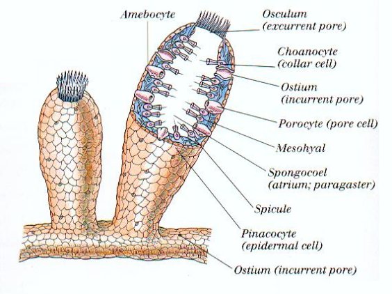
  2. Body form- body is porous. pores are of two types : inhalent pores- ostia and exhalent pores- Oscula.
  3. Symmetry- Mostly asymmetrical. Some shows radial symmetry.
  4. Germ layers- Sponges are first multicellular diploblastic animals i.e., derived from two germ layers ectoderm and endoderm.
  5. Level of organisation - have cellular level of organisation.
  6. Body wall- Body wall of common sponge consists of three layers.  
      A) Pinacoderm( dermal layer) Outer cellular layer  consisting pinacocytes and porocytes.
       B) Choanoderm( gastral layer) - Inner cellular layer which  consists of highly specialised cells called choanocytes or collar cells.
       C) Mesohyl layer ( Mesenchyme)- Non-cellular layer found inbetween pinacoderm and choanoderm. It has fine dispersed spongin fibres, spicules and amoebocytes.
7. Canal System- Three types of canal system are found in sponges: (i) Ascanoid canal system- simplest type found in Leucosolenia and few other sponges. (ii) Syconoid canal system- More complex then Ascon type. found in Sycon and some other sponges. (iii) Leuconoid canal system- most complex canal system found in spongilla.
Central body cavity of a sponge is called spongocoel or paragastric cavity.
The canal system helps the sponge in nutrition, respiration , exretion and reproduction.
8. Skeleton- Almost all sponges possess an internal skeletal system. It may consist of calcereous spicules or fine spongin fibres or of both , located in the mesohyl layer.
9. Digestion- intacellular and takes place inside food vacuoles.
10. Circulation- Distribution of food from the ingesting cells to the others is brought about by the wandering amoebocytes of mesohyl layer.
11. Respiration-exchange of gases by diffusion through the plasma membranes of the cells .
12. Excretion- By diffusion through the plasma membrane of cells. Chief excretory product is Ammonia.
13. Reproduction- Both sexual and asexual type of reproduction occurs. Asexual reproduction takes place by budding and gemmules. Sponges are hermaphrodite. Fertilization is internal.
14. Development- Zygote undergoes holoblastic cleavage. Development includes a free swimming larva, the amphiblastula( in sycon ) or parenchymula ( In Leucosolenia ) for dispersal of the species.

Unique features of phylum porifera-
Presence of Ostia and oscula , canal system and skeleton system made up of spongin fibres and spicules.

Classification
On the basis of skeleton, phylum Porifera is divided into three classes-
Class 1- Calcarea ( calcis= lime ) -The skeleton is of calcerous spicules.Examples;- Leucosolenia, sycon, grantia.
Class 2 - Hexactinellida ( hexa - six ). The skeleton is of siliceous spicules which have six rays. Ex.- euplectella, hyalonema.
Class 3- Demospongiae ( demas- frame ) - The skeleton is of spongin fibres with siliceous spicules or may be absent. Ex;- euspongia , spongilla, cliona, chalina.


  •  Leucosolenia- simplest colonial sponge consisting of a number of horizontal and vertical tubes. The development is with a larva, the parenchymula.
  • Sycon ( Scypha)- development is with a larva, the amphiblastula.
  • Euplectella( The Venus flower basket) - found in deep sea water. They are abundent near the Phillipine Island and West Indies. Its skeleton is costly marriage gift in Japan as it is thought to be symbol of union of wife and husband.
  • Hyalonema( The glass rope sponge) - It is fixed in the mud by a root tuft of long, twisted spicules. The upper surface has the gastral cone which bears opening of the excurrent canals. 
  • Euspongia - the bath sponge - surface is raised into small projections, called connules. Depressions between connules bears small pores , the dermal ostia. Its skeleton is used for bathing, washing automobiles, cleaning furniture and window panes. It is used for applying cosmetics, mopping etc.
  • Spongilla- fresh water sponge-Spongilla a common, widely distributed fresh water sponge shows various shades of green colour because of the symbiotic algae present inside.
  • Cliona - the boring sponge - The sponge can penetrate the rocks and break them into pieces  . The sponge can also bore through the oyester shell and, therefore, harmful for pearl oyester industry.
  • Chalina - the deadman`s fingers or the Meramaid`s gloves- It is shaped like a hand with several fingers perforated with oscula hence called so.

Saturday, October 7, 2017

Origin of Life and Evolution

 Read Evolution notes in pdf using link mentioned below and if you find them useful then also do share with your friends
https://drive.google.com/file/d/0B67erinnvE6LMXlNR3RidUJHTVU/view?usp=sharing
 Join our google plus community for more updates and question bank link mentioned below
 https://plus.google.com/communities/115473863387563726333
 Also do like and share our facebook page
www.facebook.com/pcbnote


Wednesday, September 20, 2017

Thermodynamics notes (Chemistry)

High quality thermodynamics notes for neet, aiims.
Thanks for showing so much interest in our notes. We will upload complete syllabus notes very soon.
Join our facebook group and follow our blog n keep sharing.


Tuesday, September 19, 2017

Applications of biotechnology (biology)

Applications of biotechnology high quality notes in pdf .
For more content stay Tuned.
Do share,follow our blog  and also help others.
For question bank like our facebook page pcb notes and join our group.