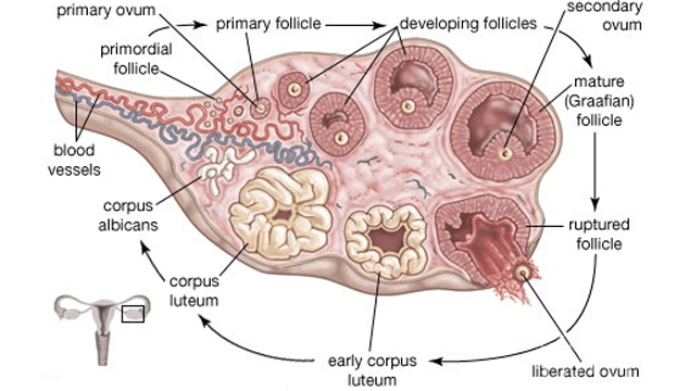
- Consists of pair of ovaries, a pair of oviducts(Fallopian tubes), a sac-like uterus and the external genitalia like vulva and genital glands.
Ovaries
- Almond shaped structures in the upper part of the pelvis. It lies in the lower part of abdomen attached to dorsal wall by mesovarium near kidney. Attached to uterus by ovarian ligament.
- Each ovary measures about 3cm*2cm*1cm surrounded by the fold of peritonium.
- Blood vessels and nerves enters through a narrow conducting part, hilus having continuity with mesovarium.
- In wall of ovary visceral peritonium, germinal epithelium and tunica albuginea are present. Germinal epithelium is made up of Primary Germ Cells(PGC) .
- Ground part of ovary is stroma differentiated into outer cortex and inner medulla , mesodermal in origin.
- P.G.C. of germinal epithelium are endodermal in origin.
- First of all from P.G.C., follicle cells are formed which fuse to form egg nest in which one is developing ovum, rest of the cells are destroyed to give nourishment to developing ovum.
- Destroyed cells atractic cells. The phenomenon is called atrasia.
- First it is single layered termed as primary follicle. Soon it becomes double layered i.e., secondary follicle after that it is converted into graffian follicle or mature follicle.
- Its formation is folliculogenesis under control of FSH and LH.
Accessory Organs
- The oviduct, uterus, vagina , clitoris, the accessory glands and mammary glands constitutes the secondary sexual organs of human female.
Fallopian Tubes/Oviducts
Fallopian tubes are a pair of muscular tubes(10cm) that extend laterally from the uterus on either side of the wall of pelvis. Fertilization occurs into it.
It is supported by double fold of peritonium.
It shows 4 regions-
(i) Infundibulum-
- Infundibulum has broad, funnel shaped proximal part.
- Its margin bears fimbriae which has finger like projections.
- In funnel ostium aperture present, from it eggs enter into duct.
- Ampulla is a thin walled, longest part lined with ciliated epithelium and is the site for fertilization.
- Very short, narrow thick walled straight part.
- It is a narrow part that communicates with uterine cavity.
- Tubectomy (or Tuballigation , the cutting or ligation of fallopian tube prevents the entry of ovum into it.
Uterus(Womb)
- For viviparous development in mammal it makes the house of embryo.
- In woman it is sac-like, thick walled, pear shaped, muscular and glandular part of 8*5*2 cm in size.
- It is large, pyriform, highly elastic and development of embryo takes place into it.
- It is located above and behind the urinary bladder and is attached to body wall by mesovarium ligament.
- It shows four regions-
- Upper wide, dome shaped fundus that receives fallopian tubes.
- Cornuae the upper corners where the oviducts enter into the uterus.
- Middle large body or corpus which is the main part.
- Lower narrow cervix that projects into the vagina.
- In the wall of uterus outer layer is perimetrium, middle layer is myometrium and inner is endometrium.
- Myometrium consists of inner and outer of longitudinal smooth muscles and middle circular muscles. Longest involuntary muscles present here.
- Endometrium consists of inner epithelial layer and underlying connective tissue layer(lamina properia) with many coiled tubular glands and screw-like blood vessels. This undergoes cyclic changes during menstruation.
- Cervix has two openings, os internalis towards corpus and os externalis towards vagina.
- Cervix communicates with uterus by internal os. and with vagina by external os.
- Cavity of cervix between external and internal os. is cervical canal.
- Cervix is composed of the biggest and most powerful sphincter muscles in the body It is strong enough to hold about 7 kg of tissue fluid.
- After implantation and pregnancy placenta is formed by the contribution of uterine wall.
- Hysterectomy is the surgical removal of uterus due to defect.
 Vagina
- A canal below uterus opens to outside between urethral pore and anus or in the urino-genital sinus(rat).
- Median, elastic, muscular tube 7.5 cm long. Opens into vestibule by vaginal orifice.
- It receives penis during copulation and also serves as birth canal.
- Space between vaginal wall and cervix is fornix.
- Initially its opening remains covered with a membranous hymen which ruptures either during sexual intercourse or due to any other vigorous physical exercise or work.
- A slit in hymen allows menstrual flow to pass out.
External Genitalia
- The external part around the vaginal pore is vulva with upper pad-like part, mons pubis with hairs.
- The middle cut the,vestibule, is laterally bound to with two pairs of lips(skin folds), outer labia majora and inner labia minora.
- At the anterior junction of labia minora a small erectile clitoris present, homologous to penis.
- In the vestibule the urethral opening is present on the upper side and vaginal orifice at the lower side.
- Area between fourchette and anus is perineum.
Glands
Vestibular glands are of two types:
- Lesser Vestibular glands/Paraurethral glands/Glands of skene: Numerous minute glands that are present around the urethral orifice. These glands are homologous to the male prostrate and secrete mucous.
- Greater Vestibular glands/Bartholin's glands: Paired glands situated one on each side of the vaginal orifice. These glands are homologous to bulbourethral glands/ cowper's glands of male and secrete viscous fluid that supplements lubrication during sexual intercourse.
Breasts
- One pair, their development is under control of pituitary.
- Nipples present (absent in prototheria). Around nipples area mammary glands present, in it melanin is maximum.
- Milk is synthesized by lactogenesis under control of prolactin. Milk is secreted under control of oxytocin.
- Early milk is rich in minerals i.e. colustrum.
- Mammary glands are modified sweat glands. Glands open on the nipples by lactiferous ducts.
- Just under nipple, lactiferous sinuses present to store milk.
Onset of Puberty in Female
Puberty attained at the age of 13.
It includes-
- Growth of the breasts .
- Growth of external genitalia.
- Broadening of pelvis.
- Growth of pubic hair.
- Increase in sub cutaneous fat.
- Starting of mestrual cycle i.e. Menarche and stoppage of menstrual cycle is known as Menopause
Histology of Ovary
- Covered with connective tissue sheath tunica albuginea , its inner regions are distinct as outer cortex and inner medulla.
- Cortex consists of reticular fibres, spindle shaped cells and ovarian follicles;medulla has stroma.
- Along the germinal epithelium groups of oogonia (egg nest) project into cortical part as egg tubes of Pfluger , which give rise to ovarian follicles.
- About 5 lac primary follicles are formed only once when female fetus is only three months old in her mother`s womb. Follicles remain arrested in diplotene stage of prophase-I and become active only after 10-12 years of age for maturation influenced by FSH.
- Maturing follicles secrete estrogen that maintains the sex organs and secondary sexual characters.
- In human only one follicle releases ovum each month.
- Majority of follicles degenerate in the process and called atretic follicles or atresia.
Graffian follicle
- It appears as Knob or stigma in medulla.
- It is round, discovered by Graff.
- It is covered by two layers - theca externa and theca interna.
- From theca interna estrogen is secreted to control secondary sexual characters of female.
- Inside theca theca interna, membrana granulosa is present.
- Secondary oocyte is attached to covering layer by group of cells i.e., discus proligerous.
- The point of discus proligerous close to secondary oocyte is Hill of follicular cells or Cumulus oophorous.
- Jelly-like covering around the oocyte is zona pellucida , a primary membrane secreted by egg itself.
- The layer of granulosa cells around the oocyte is called corona radiata for having radiating process to draw nutrients from liquor folliculi.
- Cavity of follicle is antrum or follicular cavity filled with follicular fluid.
- Graffian`s follicle is ruptured, secondary oocyte comes out of ovary. It is ovulation and at this time L.H. is secreted more.
Corpus Luteum
- The ruptured graafian follicle after ovulation forms separate glandular structure with yellow pigment called as corpus luteum (yellow body) under the influence of LH.
- It secretes a little amount of estrogen and mainly progesterone hormone that maintains pregnancy. It develops extensively if pregnancy occurs, otherwise, degenerates to form corpus albicans (white body).
- Corpus luteum secretes progesterone and relaxin.
- Progesterone is helpful in implantation, to stop ovulation during gestation period.
- Relaxin is harmful to relax pelvic muscles during partition.
- 1.5 cm in diameter, reaching the stage of development 7-8 days after ovulation. Then it begins to involute and eventually loses its secretory function as well as its yellowish, lipid characteristic about 12 days after ovulation, becoming then the so called corpus albicans.
Subscribe to our YouTube channel for video lectures


No comments:
Post a Comment